
Glaucoma Consults Round-up: Pt 1. Sep 28 - Oct 2, 2009 w/HRT and VF results
/A recap of this past week's tweets about patients referred to me for glaucoma management. This article contains additional information not found in the original 140 character postings. Please add your comments so that we can extend these teachable moments over time.
Wednesday September 30, 2009
Case 1:
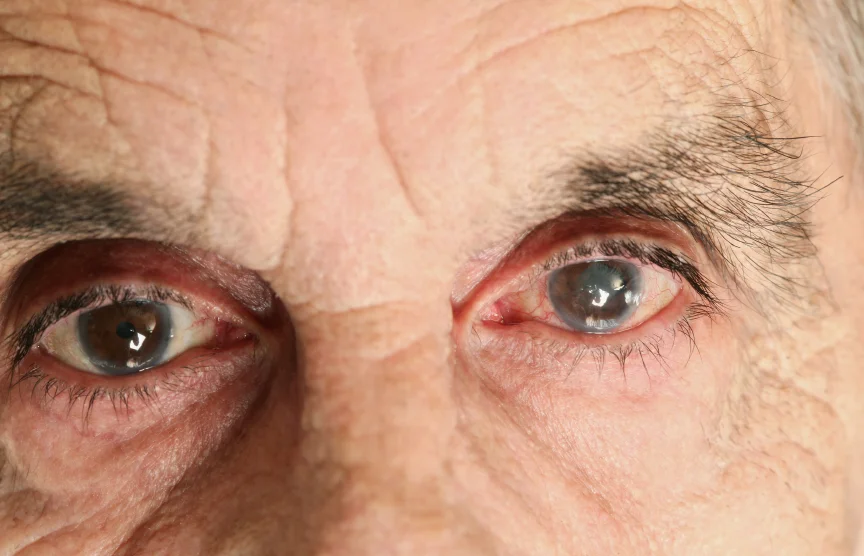
robschertzer #glaucoma #consult 59 yo WF, disc asymm, +ve FamHx, thin CCT, IOP 18,20; will follow over time to see if progresses
This patient had been receiving HRT nerve head scans at my office as ordered by the referring optometrist every 6 months for the past 1.5 years. Although not showing signs of progression in that time, given her family history of glaucoma and her suspicious appearing optic nerves, she was referred to see me. She is a 6 D myope with 'increased cupping' of the left optic nerve compared with the right, tilted optic nerves with peripapillary atrophy (from the myopia.) Central corneal thickness measurements were a bit thin at 509 and 501 ums with IOP readings of 18 and 20 at 1300hrs.
Case 2:
robschertzer #glaucoma #consult 9 yo Asian M, physiologic cupping vs glaucoma?, mod myope, HRT GPS 70%; looks physiologic, will follow annually
30Sep2009 Case 2 HRT Stereo
On rare occasion, I am referred children as glaucoma suspects, usually on the basis of 'increased cupping.' (I'll explain the parenthetical use of cupping in a future article.) For this young man, the Visual Acuity was 6/6 in both eyes with his -3.5D correction, visual field testing was normal, no abnormalities on visual field testing and a baseline HRT nerve scan was performed for future comparison.
Commonly with physiological 'increased cupping,' we see that disc area is larger than normal. That is, the overall size of the optic nerve. Therefore, even though the cup area is well above average, there is still a normal amount of healthy optic nerve tissue as reflected by the rim area value above.
Case 3:
robschertzer #glaucoma #consult 67 yo E Indian M, optom referral ?notch optic nerve; confirmed paracentral VF defect & notch; awesome job by optom!
30Sep2009 VF OS
This patient was referred by his Optometrist for increased optic nerve 'cupping,' and abnormalities on Visual Field testing. The referring optom was also concerned that the inferior rim of the optic nerve was quite thin/notched from glaucomatous damage. The patient's medical history was significant for high blood pressure and diabetes.
Visual acuity was 6/6 in each eye, IOP readings 16 mmHg OD and 18 OS, there were lens implants in both eyes from prior cataract surgery, drainage angles were wide open and the inferior rim of the left optic nerve was indeed notched. Visual Field testing correlated with the damage to the inferior rim by showing a defect in the paracentral superior field of vision in the left eye.
Case 4:
robschertzer #glaucoma #consult 33 yo Asian M, 3D myope, increased cupping, distant relative glaucoma; will follow 2 yrs.
Again a patient referred for 'increased cupping.' He had been referred to me 5 years earlier as well at which time I was not terribly convinced of a significant glaucoma risk. Visual Acuity was 6/6 OU with current -3.25 D correction, IOP 13 mmHg OU at 1410hrs, wide open angles and physiologic cupping, with normal visual field testing. Opting to see patient in a couple of years for follow-up.
Case 5:
robschertzer #glaucoma #consult 30 yo Asian F, strong fam Hx glaucoma/blindness, prior LASIK so thin CCT, IOP 11, tilted discs; follow for now
This patient has a strong family history of glaucoma effecting, maternal grandmother, aunt, uncle as well as her mother and was noted by her optometrist to have 'increased cupping' of her optic nerves. Visual Acuity was 6/7.5 OD and 6/6 OS with unreliable IOP readings (due to surgically altered corneas) of 11 OU at 1450hrs. As with many myopes, despite refractive surgery as this does not change the optic nerve, her optic nerves are tilted and show peripapillary atrophy, both of which are risk factors for progressive glaucoma (not to mention her strong family history.) She will require regular re-assesments every 4-6 months to rule-out progressive damage to her optic nerves from developing glaucoma. She is being observed for now without treatment.
Case 6:
robschertzer #glaucoma #consult 61 yo WF, suspect b/o VF, IOP 16 OU, v small cups; mostly learning curve, will repeat VF
30Sep2009 Case 6 VF OD
This patient was referred with a visual field test performed at the referring Optometrist's office as shown below. Optic nerves were very healthy in appearance with 0.3 cup-to-disc ratios in both eyes, IOP readings of 16 OU, wide open angles. They were also noted to have significant blepharitis. The patient was started on warm compresses and lid hygiene for the blepharitis and will return in 3 months for repeat testing until she overcomes the 'learning curve' for visual field testing. Some patients need to repeat the test several times until they get better at performing it.
Thursday October 1, 2009
Case 7:
robschertzer #glaucoma #consult 65 yo F, prior LPI, gonioplasty, Pilo & Cosopt controlled plateau iris; IOP now 46 & 60! +diamox, lumigan see few days.
This is one of the scariest cases of recent times for me and is the subject of a separate article on my site as well to bring it to the attention of more people. I have been following her for more than 3 years for Plateau Iris Syndrome which has been stable following SLT, LPI and Gonioplasty combined with taking Pilocarpine drops in both eyes and Cosopt in the right eye.
For some reason though, she showed up for her regular visit today noting blurry vision, though measuring 6/7.5 OD and 6/9 OS with her current spectacle correction. Her IOP readings were almost off the scale at 46 OD and 60 OS @0930hrs. The only other finding of note was that the pupils were mid-dilated.
Laser and medical treatment of plateau iris syndrome can be quite frustrating; IOP can be fine then be high at a future visit. It was somewhat disappointing for physician and patient alike to have this high an eye pressure all of a sudden. I added Diamox and Lumigan, asked her to use the Cosopt in both eyes instead of just in the right, and to keep using the Pilocarpine in both eyes.
An hour later I received a call from her pharmacy. They were wondering if the fact that they gave her homatropine instead of pilocarpine when she refilled her prescription in March could have effected her pressure reading?! Our records and those of the pharmacy clearly show the Rx was for pilocarpine but they gave her homatropine which they then cover completely with their prescription label. However, the red bottle cap was what tipped off the pharmacist of their error. The patient did not bring her drops to her visit but did bring them to her pharmacy. Later that day, the pharmacist sent a CYA type fax stating that the bottle they mistakenly gave her in March was almost completely full so that it is unlikely she used much of it.
I cannot even begin to express my concern over this pharmacy dispensing error and how it may have resulted in permanently damaging this patient's vision. Time will tell in follow-up whether the damage they caused can be reversed.
1Oct2009 Case 7 Note from Pharmacist
Case 8:
robschertzer #glaucoma #consult 48 yo Asian M bungee cord injury IOP 36 max meds; booking for Trab MMC.
After that lost shocker, this patient was relatively more straightforward in terms of management. After striking his left eye with a bungee cord one month prior to seeing me, his eye pressure was 36 taking DuoTrav, Alphagan, and diamox. Despite the large eye pressure asymmetry (IOP just 16 in the fellow eye) he is bound to begin to show progressive damage from glaucoma if left untreated.
Friday October 2, 2009
Six more glaucoma consults and already wrote enough in this space so look for Part 2 of this past week's glaucoma consults to be posted soon.