Glaucoma Consults Round-up: Nerve fibre imaging, herbal treatments, and rule-out brain tumour!
/Friday October 9, 2009 & October 28, 2012
This was one of many articles corrupted during the migration from Squarespace version 5 to version 6. I have now manually restored the article and hope that it is still of some value.
Case 1:
robschertzer #glaucoma #consult 49 yo WM Mom w/ COAG, N exam except v dark pigmented TM, no signs PDS or PXF. Form fruste of either?
Referred by optometrist with IOP readings of 23 OD and 24 OS @1050hrs and whose mother is known to have Chronic Open Angle Glaucoma (COAG.) When seen by me, his VA was 6/7.5 OU, IOP readings 20 OU @09040hrs and angles were wide open. Optic nerves appeared normal as were HRT and VF tests. An annual check should be sufficient for this patient as no findings aside from the family history and age > 40 yo were present.
9Oct2009 Case 1 HRT OU
9Oct2009 Case 1 VF OS
9Oct2009 Case 1 VF OD
Case 2:
robschertzer #glaucoma #consult
42 yo WF disc asymmetry, N VF & CCT, remote FamHx glaucoma, abn FDT not confirmable by SAP.
Referred with asymmetric optic nerves such that the left optic nerve appeared to have more 'cupping' than the right without a corresponding IOP asymmetry. Visual acuity found to be 6/6 OU with current correction and IOP of 16 OD and 14 OS at 0940hrs. Anterior segment findings were all normal and optic nerve findings as per referral with slight tilt to both optic nerves and a thinner inferior rim to the left optic nerve. Patient will be seen in 6/12 to re-assess with both tests repeated at that time.
9Oct2009 Case 2 HRT GPS OS
9Oct2009 Case 2 VF OS
9Oct2009 Case 2 VF OD
Case 3:
robschertzer #glaucoma #consult 68 yo WM, blinded by glaucoma OS and soon to be OD; refusing Tx x yrs in favour of herbal remedies
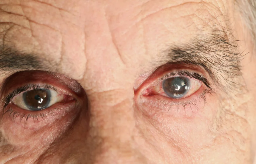
This patient presents as an exercise in frustration. Yes, there is a role for herbal therapy in glaucoma but it usually cannot replace modern treatments completely. This patient firmly believes it can, even though he has been blinded in the left eye by ignoring all medical advice for many years before coming for a second opinion. He provides his own proof that herbal remedies alone are inadequate by having wiped out his left optic nerve completely by sticking to his beliefs.
He was originally diagnosed with glaucoma in 1986 in another province, started on drops for glaucoma but stopped them because his pressure went down. Followed by a local ophthalmologist since 1997, he has been referred to a glaucoma colleague of mine in town a couple of times over the years 1997. At that time, his eye pressures were documented as 25 and 26, his right optic nerve was completely healthy and his left optic nerve showed significant damage but the rims were still intact. His records are quite lengthy and include a letter written to the patient expressing concern over his failure to take recommended glaucoma therapy in 2005. There is also clear documentation of his continued loss of visual field relentlessly from 1997 until 2009.
9Oct2009 Case 3 HRT GPS OU
9Oct2009 Case 3 VF OS
The findings in our office were visual acuity of 6/6 in the right eye and Count Fingers at 3' in the left. IOP readings were 21 mmHg OD and 22 OS at 1010hrs. His angles were open for 360 degrees to the posterior trabecular meshwork. The HRT nerve scan above adequately correlates with the clinical findings of advanced glaucomatous damage to both eyes. His visual filed illustrates the marked damage as well to the left eye and impending doom to his right.
The resident currently on our glaucoma service and I spent a lot of time with this patient. We discussed how herbal treatments can be complementary to our western medicine but are not adequate on their own. In fact, the patient has managed to have his vision pretty much wiped out by relying only on herbal therapy for much of the past 23 years and is on the verge of wiping out his right eye as well. We gave our pitch for using a topical medication, trying just with one medication to get him on the least amount of medicine that will do the job.
We seemed to reach an understanding and he left the office with a sample and prescription for Travatan-Z. He had used a different prostaglandin analogue before that bothered him so we chose this as one likely to be well tolerated because of not having the benzalkonium chloride common to many other drops and still very effective with once daily dosing. He was instructed to stop at the front desk on his way out to book his pressure check for 4-6 weeks from now; he left without booking the appointment. I suspect we will never see him again even though we will leave messages for him.
Case 4:
robschertzer #glaucoma #consult 54 yo WM abN OCT by referring optom; not useful test modality for glaucoma as no account nerve diameter
Is an abnormal Ocular Coherence Tomography (OCT) result enough of a reason to suspect glaucoma? This patient is a -8.50D myope with IOP readings of 12 OU at 1700hrs by the referring optometrist and was noted to have an abnormal OCT using the 'optic disc cube 200x200' strategy as illustrated below along with the Heidelberg Retinal Tomogram (HRT) done in my office.
9Oct2009 Case 4 OCT
9Oct2009 Case 4 HRT GPS OU
Comparing the two modalities for this patient, note the red ring drawn around the optic nerve in the RNFL Thickness Deviation image on the OCT printout; this is the 2nd row of 4 rows of images. Compare this with the green line outlining the optic nerve in the 2nd row of the HRT scan. These lines represent where these devices are measuring the thickness of the nerve fibre layer. Note that this is almost a full optic nerve diameter in distance from the edge of the optic nerve on the OCT whereas it is the outer edge of the optic nerve in the HRT. The HRT always uses the optic nerve periphery for determining the nerve fibre layer thickness, regardless the size of the optic nerve. The OCT, on the other hand, can be thought of as applying a cookie cutter centred on the optic nerve to determine where it will measure the nerve fibre layer thickness. For normal or small optic nerves, this distance is nowhere near the edge of the optic nerve and for large optic nerves, this can be part of the nerve itself and not even be a measurement of the nerve fibre layer at all.
9Oct2009 Case 4 VF OS
9Oct2009 Case 4 VF OD
When reviewing the visual field tests done at the referring optometrist's office, the Pattern Deviation plot looks completely normal. This cleans up the Total Deviation plot by subtracting the 7th highest threshold value from all the points in order to reveal any patterns of visual field loss such as nerve fibre bundle defects that might be hidden if everything looked black as it does on the total deviation for this patient. This is usually meant to correct for media opacities such as a significant cataract. This patient however does not have any significant cataract formation so the decreased sensitivity could actually be early glaucoma damage showing as overall decreased sensitivity to the lights without any specific nerve fibre bundle pattern. Decreased sensitivity is a common finding in glaucoma even though it is not specific to glaucoma.
Overall it was decided that this patient is a 'glaucoma suspect' on the basis of these findings but we could not confirm glaucoma. They will return in 6 months at which time the referring optometrist will have already repeated the VF and we will repeat the HRT nerve scan.
Case 5:
robschertzer #glaucoma #consult 80 yo WF w/ ARMD, had IOP 31 by tonopen referring retina doc; no signs glaucoma at all
9Oct2009 Case 5 HRT GPS OU
9Oct2009 Case 5 VF OS
9Oct2009 Case 5 VF OD
This patient has been followed by the retina service for macular degeneration and usually runs IOP readings in the low to mid 20s, always higher in the left than the right. When the IOP was found to be 31 in the left eye recently, she was referred for this glaucoma assessment. This patient's VF defects are related to her macular degeneration with no evidence of any increased risk of glaucoma. IOP readings were 13 OD and 18 OS with CCT of 539 and 553 ums. Nerves look healthy. No follow-up with me has been arranged as she will keep seeing her retinal specialist.
Case 6:
robschertzer #glaucoma #consult 69 yo WM, noted loss periph vision OS so got checked; IOP 10 OU, CCT 665!, OS disc cupped beyond nerve.
9Oct2009 Case 6 HRT Stereo OS
9Oct2009 Case 6 VF OS
This wins the award for biggest conundrum of the week; I guess I would say that on the rare occasions that we need to make sure a patient doesn't have a brain tumour! This patient had last been seen by an Optometrist 10 years earlier before returning to see one recently when he noticed a piece of his vision to be missing in the left eye. There were apparently notes about an abnormal looking optic nerve but nothing more specific in the old records.
IOP from Optom was 18 OD and 22 OS by Non-Contact Tonometry, VA 6/12 OD and 6/15 OS with mixed astigmatic correction.
When examined in our office, his IOP readings were just 10 OU using Goldmann applanation tonometry and his CCT measurements were a remarkably thick 665 and 661 ums. Remarkable as we often associated HIGH IOP readings with thick corneas which can give a falsely elevated reading since the 'gold standard' tonometer, the Goldmann Tonometer, depends on indenting an eye of a standard thickness. His left optic nerve showed extensive 'cupping' with extensive atrophy surrounding it and a greatly thinned nerve fibre layer. One would expect this to just be a congenital anomaly, rather than acquired over time from glaucoma damage, especially given such extremely low eye pressures in the presence of thick corneas.
We arranged for him to get a Goldmann Visual Field, as opposed to the more common Standard Automated Perimetry testing. Rather than testing 'static points,' the Goldmann is a 'kinetic' test in which the target lights are moved from the far periphery inward until they are seen. In addition, rather than a single sized target, the size of these lights varies.This gives a more refined mapping of the field of vision than any automated perimeter can achieve but requires specially skilled technicians to perform the test and more time. We are specifically looking for the presence of a 'junctional scotoma.' This would be a central defect in one eye and a 'pie in the sky' in the fellow eye, that would go along with a tumour behind the left eye near the area of the optic nerve chiasm such that it effects the right eye. There could also be other neurological visual field defects that could also help localize a lesion given that there are no other neurological complaints in this patient.
When this patient returns, we will be able to decide if he needs any special imaging of his brain.
Case 7:
robschertzer #glaucoma #consult 69 yo WM on Vitalux for ARMD, inf arcuate defect; confirmed Glaucoma too, pt averse to drops so SLT.
This patient was referred by his Optometrist with increased optic nerve 'cupping,' abnormal Visual Field, thin corneas. IOP readings were 22 OD and 23 OS @1045hrs with VA 6/9 OD and 6/12 OS. This patient was so adverse to using eye drops that it was even hard to get the topical anasethesia drops in his eyes to check his eye pressures. Although this will make it a challenge to perform SLT laser, this is a better option for him than expecting him to take glaucoma drops.
9Oct2009 Case 7 HRT GPS OD
9Oct2009 Case 7 HRT GPS OS
9Oct2009 Case 7 VF OS

