Glaucoma Consults Round-up - Toxic beta blockers, red hot eyes and more
/A recap of this past week's tweets about patients referred to me for glaucoma management. Topics discussed include systemic and ocular risks of excessive use of medications. This article contains additional information not found in the original 140 character postings. Please add your comments so that we can extend these teachable moments over time.
Monday October 5, 2009 & October 28, 2012
Another article restored meticulously after disaster struck. Enjoy!
Case 1:
robschertzer #glaucoma #consult 67 yo WF uveitic glaucoma on every med and also had LPI? OD. IOP 34 OD 19 OS, VA CF OD 6/120 OS. Needs TrabMMC.
Urgently referred from out of town for intractable glaucoma in a patient who is almost blind after bilateral vitrectomy and diabetic laser treatments. Referred to see both me and a retina surgeon while in town. Patient is already taking Combigan, Xalacaom, Alphagan, Azopt and Cosopt drops and Diamox pills for glaucoma as was well as Voltaren drops for inflammation.
It seems to happen that patients at times get placed on multiple medications for glaucoma, as they likely do for other medical conditions. At times it seems that there is either something lost in communication with the patient or panic from the doctors that precludes a more systematic approach to treatment. This patient is on three different glaucoma combination drops that each contain timolol in them (Combigan, Xalacom, and Cosopt) and is also on a systemic beta blocker (atenolol) which may already be giving her a fully beta blocker response making topical application useless. All these beta blockers are likely to land her in hospital or 6 feet underground by dropping her blood pressure too low, blunting any hypoglycemic response from her diabetes, not to mention the toxicity of extra eyedrops to the surface of the eye. This patient is also on two different topical carbonic anhydrase inhibitors (CAIs) - Azopt and Cosopt, and an oral CAI - Diamox. High concentrations of topical CAIs are required to fully block the enzyme in this pathway, so sometimes supplementing one topical CAI with an oral CAI can help reach a better response; there should not be a need though for two different topical CAIs.
This patient will be having a trabeculectomy in the near future so we will be able to get her off all her meds, at least temporarily and hopefully longterm. In the meantime, we need to minimize her exposure to toxic levels of beta blockers and the preservatives contained in all these redundant eye drops that just serve to cause side effects with no therapeutic benefit.
Wednesday October 7, 2009
Case 2:
robschertzer #glaucoma #consult 26 you Asian M inc vertical cupping OD>OS, N CCT, mom followed for glaucoma, IOP 12 OU @1320hrs. See in 2 yrs.
This patient has increased 'cupping' of his optic nerves and his mother is being followed for glaucoma. The main teaching point as often comes up is whether this appearance of increased cupping of the optic nerve represents any loss of healthy neuroretinal rim tissue or if the optic nerve's rim tissue still contains the approximately 1 million axons coming from the retinal ganglion cells.
7Oct2009 HRT Stereo OS
7Oct2009 Case 2 HRT Stereo Disc simulated view OS
Looking at the stereometric parameters, the disc area is larger than normal as is the cup area, but the rim area is at the far upper end of normal as well. The Glaucoma Probability Score looks at a mathematical model of the 3-D structure of the patient's optic nerve to compare that with patients known to have glaucoma and gives a probability score in which 0.8 would mean 80% chance of this patient having glaucoma. For this patient, the GPS score is suspiscious enough to certainly keep following this patient to see if they develop changes in the shape of their optic nerve on follow-up testing or in the function of the nerve as shown by Visual Field testing.
Case 3:
robschertzer #glaucoma #consult 63 yo phillipino F very shallow ant chambers but angles still open to post TM; dense cataracts will be removed as Tx
This patient has been seen for HRT nerve scans for the past year or so but has now been referred to see me as a glaucoma suspect on the basis of increased vertical cupping of her left optic nerve which corresponds with a nasal step on Frequency Doubling Technology (FDT) perimetry. No copy of the FDT was provided but that point is somewhat moot as there are too many false positive results in the FDT screening test to make it terribly useful. As a screening tool though, it is better to over-call possible glaucoma than to miss obvious glaucoma damage so this test does have some value. However, the optometrist did the important next step of following up the screening FDT with Standard Automated Perimetry (Medmont or Humphrey both fine) for threshold determination of each point. Threshold testing means determining the dimmest light the patient sees at each test point by presenting stimuli of varying intensities until the value is determined within a narrow range of +/- 2 DB.
Case 4:
robschertzer #glaucoma #consult 59 yo E Indian M, COAG, prior SLT OU, multi topical meds, IOP varies 15-25, VF confirm worsening; needs Trab MMC
This patient was referred by a comprehensive ophthalmologist who has followed him for the past 5 years. On presentation at that time, he had IOP readings around 20 and a 'fairly damaged' right optic nerve. Eye pressures have remained around 20 with drops and more recently a Selective Laser Trabeculoplasty (SLT) laser but recently after raised to the mid 20's. The referring ophthalmologist is concerned that they will continue to progress at this pressure level.
Current medications for this patient included Timolol and Lumigan. Of note again with this patient is that he is already also on a systemic beta blocker so that he may already have a maximal IOP lowering effect from that oral agent such that the timolol may be adding more side effects than pressure lowering. Visual acuity was quite good at 6/9 OD and 6/7.5 OS with IOP readings in our office of 15mmHg OU @1350hrs. Angles were wide open and optic nerves showed advanced damage, including collapse of supratemporal rim OS and superior and inferior rims OD. The VFs matched the optic nerve findings with a nasal step OS and inferior and superior defects OD.
Although somewhat fuzzy in terms of the image quality and also SITA-FAST instead of SITA-STANDARD was used to decrease the accuracy of the test, a faxed copy of the patient's VF tests were provided by the referring doctor. These VFs both confirm progression in the right eye more than the left despite today's IOP reading. I rarely recommend sacrificing visual field quality by skimping on the testing strategy. The FAST is a less precise test than the STANDARD in terms of how closely it determines the threshold value for each point. The STANDARD testing strategy uses a 'double-bracketing' technique vs a 'single-bracketing' technique for the FAST test. Double-bracketing refers to narrowing down the dimmest light that can be seen at any given point by first crudely determining the value with 4dB steps in light intensity and then when bracketed between too bright and too dim, the threshold is then narrowed down with 2dB steps. In the FAST test, the threshold is only crossed once, using 3dB steps. The FAST technique is therefore not as precise in determining that threshold light intensity that is perceived at every point on the exam.
7Oct2009 Case 4 VF GPA OS
(Click on the Glaucoma Probabilty Analysis VF images above in order to enlarge them.)As you can see, there are quite a few completely filled triangles; these indicate areas on the VF that have changed for the worse on 3 or more consecutive visits. This is considered definite progression that warrants a change in treatment. In this case, Trabeculectomy with Mitomycin, for both eyes.
Case 5:
robschertzer #glaucoma #consult 47 yo E european, thin CCT 482/495, IOP 18/19 @1345hrs, mom w/ COAG; disc asymmetry & N VF; re-assess 6/12
This patient does have a number of risk factors that increase her risk for glaucoma, even though there is not enough evidence to support a diagnosis of glaucoma at this time. Her corneas are very thin, which is a risk factor independent of intraocular pressure for glaucoma. Below are thumbnail links to her rather normal HRT GPS results (Heidelberg Retinal Tomogram - Glaucoma Probability Score.)
7Oct2009 Case 5 HRT GPS OD
Case 6:
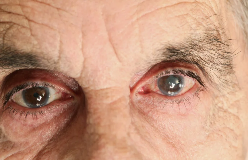
robschertzer #glaucoma #consult 36 yo E Indian M, kidney failure, horrible health, blind OD s/p sil oil PPV, eye flaming red OS mult-meds; needs Trab MMC
This patient had Acute Retinal Necrosis in both eyes that resulted in multiple retinal surgeries in the right eye with complete loss of vision (NLP.) He had a renal transplant in 1990 at 17 years of age back in India for which he is on cyclosporin along with other medications. He was referred to me with IOP of 28 in his now only seeing left eye taking Travatan, Cosopt, Alphagan as well as PredForte in his left eye for consideration of performing a trabeculectomy. His vision appeared to him to have improved shortly after his recent cataract surgery in August but has since become blurry again.
His eyes were flaming red, likely much of this from intolerance to the glaucoma medications. His cornea was moderately hazy and anterior chamber showed 4+ flare but no inflammatory cells. His IOP was only 16 mmHg at 1410 hrs. He was very difficult to examine as his eyes were very sensitive to light.
Given all his troubles that are at least in part related to irritation from his drops, I will proceed with performing a trabeculectomy with mitomycin-C. It was challenging just trying to examine him in the office so that there may be some challenges in the operating room trying to do this surgery. In addition, given his overall very poor health, it may be difficult to get clearance from anaesthesia to have surgery performed.
Case 7:
robschertzer #glaucoma #consult 84 yo WF PXF glaucoma OU, IOP 29/31, disc heme OU, compliance issues b/o memory; booking for Trab OS then OD
There seems to be no shortage of patients in need of glaucoma surgery this week. Although some of the above cases may be more controversial as to whether surgery will ultimately be their best bet, for this patient with Pseudoexfoliation Glaucoma there is documented change in visual fields, intolerance to topical beta-blockers, non-compliance to other meds and high pressures. They were referred by a medical glaucoma specialist.
Case 8:
robschertzer #glaucoma #consult 64 yo phillipino M ACG OU w/ IOP 30 OS; needs Trab MMC
This patient with was referred by the same medical glaucoma specialist with chronic angle closure glaucoma that was "suddenly uncontrolled." IOP readings were initially 25 OD and 32 OS ten years ago with severe visual field loss. Angles were narrowly opened and patient has undergone both Laser Peripheral Iridotomy (LPI) as well as Selective Laser Trabeculoplasty (SLT) in both eyes. They were also being treated upon referral to me with Lumigan, Cosopt, Alphagan as well as oral diamox.
On examination, VA was 6/9 OD and 6/12 OS with IOP readings of 16 OD and 30 OS @1450hrs. Moderately advanced cataracts were noted in both eyes and, interestingly, the left optic nerve has a healthy looking neuroretinal rim compared to the right despite having the higher eye pressure at this time. However, if we don't do something more definitive in the form of glaucoma filtration surgery, there will be significant damage to this left eye as well.

